The wide complex tachycardia (WCT) was terminated following treatment in the AED. A follow-up ECG was then performed.
Which of the following stagement is most approprite?
Choose the correct answer.
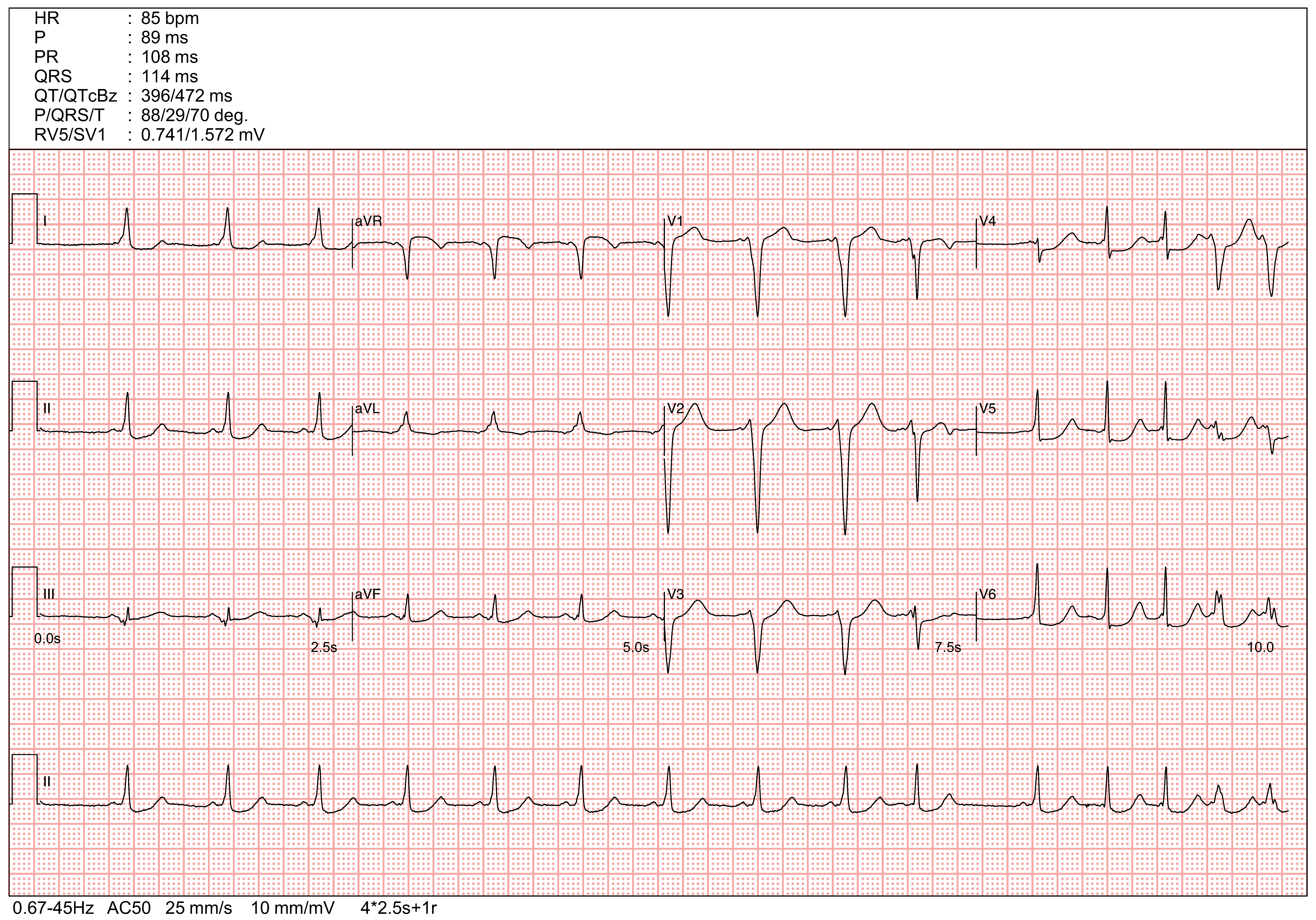
The follow up ECG shows sinus rhythm, with occasional atrial ectopics, short PR interval with delta waves.
The positive delta waves in V1 (with R<S in V1), I, II, aVF and negative delta waves in lead III are suggestive of right lateral accessory pathway.
The pre-excitation is incomplete due to fusion of electrical conduction between the AV node and the accessory pathway. In the last 2 QRS complexes, the QRS duration is widened with fully manifested pre-excitation following atrial ectopics. This is likely due to the electrical refractoriness of AV node following the atrial ectopics, allowing the electrical conduction predominantly going through the assessory pathway, which results in full pre-excitation.
The delta waves mophologies and QRS axis are similar with that in the WCT. The ECG finidngs in sinus rhythm further confirms the initial postulated diagnosis of anti-dromic AVRT mediated by a right lateral accessory pathway (most likely a atriofascicular accessory pathway/Mahaim fiber).
A is incorrect as the diagnosis is not suggestive of acute ST elevation myocardial infarction.
B is incorrect. The last 2 widen QRS complexes are likely due to atrial ectopics with fully manifested pre-excitation. The atrioventricular conduction predominatly goes through the accessory pathway rather than the AV node during the atrial ectopics when the AV node is made electrically refractory.
C is incorrect as amiodarone could block the accessory pathway and as long elimination half-life, which will make subsequent electrophysiology study/mapping and ablation of the pathway difficulty.
D is the correct answer. The patient has WPW and the previous wide complex tachycardia is likely an Antidromic Atrioventricular reentrant tachycardia (aAVRT).
E is incorrect as the diagnosis is suggestive of WPW with aAVRT.